
Address access geometry
A fine fiber and short catheter eliminate the introducer sheath for a segment in which standard access is impractical.
SHEATHLESS ACCESS · SHORT DEEP MUSCLE BRANCHES
UFLAA 400 μm fiber passes through an 18G catheter without an introducer sheath. One case involving a 1.5 cm deep muscle branch explores access in challenging anatomy.
01 / THE CLINICAL CHALLENGE
Competent saphenous trunks do not exclude pathological reflux from a deep branch.
A 45-year-old woman presented with leg heaviness, swelling and a popliteal venous aneurysm 26 years after wide resection and radiotherapy for a left-leg rhabdomyosarcoma. Dense fibrosis and scarring in the surgical field complicated access.
Duplex ultrasound showed competent great and small saphenous veins. The reflux source was a deep muscle branch only 1.5 cm long, measuring 2 mm at its origin and dilating to 5 mm near the aneurysm.
Source: Yang CC. JVS–CIT. 2026;102274. Case report, Technique, Discussion Original article
02 / THE UFLA TECHNIQUE
Understand fiber size, access and energy delivery as separate elements.
(600 − 400) ÷ 600 = 33.3%
400 μm = 0.4 mm, or two-thirds of the comparator’s diameter.
The circles are proportional to the reported 600 μm and 400 μm diameters. They are dimensional markers, not device cross-sections showing coatings or tip construction. The comparator 600 μm fiber requires at least a 16G catheter in this report; the 400 μm fiber passes through 18G. Catheter sizes are not assigned the same ratio.
Under ultrasound guidance, the target branch is cannulated. The metal stylet is removed, leaving a short plastic cannula through which the 400 μm radial fiber passes. “Sheathless” means that the introducer sheath is omitted; a peripheral intravenous catheter is still used.
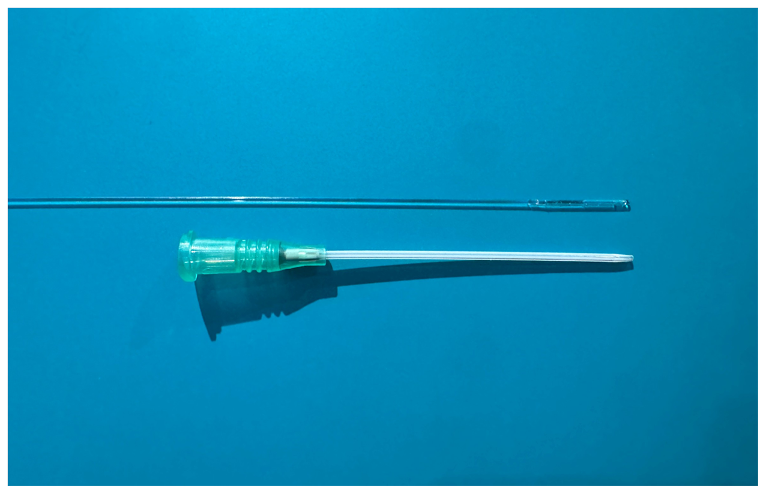
The sheathless design addresses the length occupied by an introducer sheath. Stable catheter fixation and clear ultrasound visualization of the fiber tip remain essential.
The deep muscle branch was treated at 7 W with 1-second pulses and 2-mm pullback increments (stop-and-shoot). Ultrasound verification before each emission was intended to confine delivery to the target segment.
The four positions illustrate sequence only, not the reported total number of pulses, full treatment length, a fixed pause duration or thermal spread. These are this UFLA case’s parameters; SPOT data are not substituted.
A fine fiber and short catheter eliminate the introducer sheath for a segment in which standard access is impractical.
Smaller access, position checks and tumescent anesthesia have procedural rationale. The report does not compare perforation rates, tissue temperature or nerve-injury rates.
Duplex ultrasound confirmed target-branch occlusion at three months. The case supports feasibility, without establishing comparative superiority.
Source: Yang CC. JVS–CIT. 2026;102274. Technique, Discussion Original article
03 / OBSERVATIONS & LIMITATIONS
A single case report, without a control group.
45-year-old woman · CEAP C3
Clinical and duplex assessment
Includes superficial procedures
Observation in this case
Source: Yang CC. JVS–CIT. 2026;102274. Results, Limitations Original article
04 / ASSESSMENT & CARE
Symptomatic reflux from a short deep muscle branch or perforator, especially when sheath deployment is difficult or anatomy has been altered by surgery or trauma. These are potential applications discussed in the report, not broadly validated indications.
Comprehensive duplex imaging must assess truncal competence and establish the target branch as the primary, symptomatic reflux source. The great and small saphenous veins were competent in this case; the finding cannot be generalized to all patients with truncal reflux.
Unstable catheter fixation, excessive shortness or tortuosity, or inability to continuously identify the tip on ultrasound. Limited quartz-fiber flexibility can lead to fracture or vessel injury, with off-target thermal damage or incomplete ablation.
Use clinical history, symptoms and duplex ultrasound to identify the responsible short branch and assess stable access.
In this case, prone positioning and local anesthesia were used. Ultrasound guided 18G catheter access, stylet removal, fiber placement and tumescence.
The branch was treated with 7 W, one-second pulses and 2 mm withdrawal, confirming position each time. Superficial disease received separate laser treatment and phlebectomy.
The reported three-month visit assessed symptoms and flow. Aftercare, activity and follow-up schedules remain individualized.
Source: Yang CC. JVS–CIT. 2026;102274. Technique, Discussion, Limitations Original article
RESEARCH & PUBLICATION
A 400 μm fiber passed directly through an 18G catheter to treat a 1.5 cm refluxing deep muscle branch. Occlusion was confirmed at three months in one patient. Broader applicability and long-term outcomes remain uncertain.
Version of record · August 2026, volume 12, issue 4 · Journal of Vascular Surgery Cases, Innovations and Techniques
DOI: 10.1016/j.jvscit.2026.102274
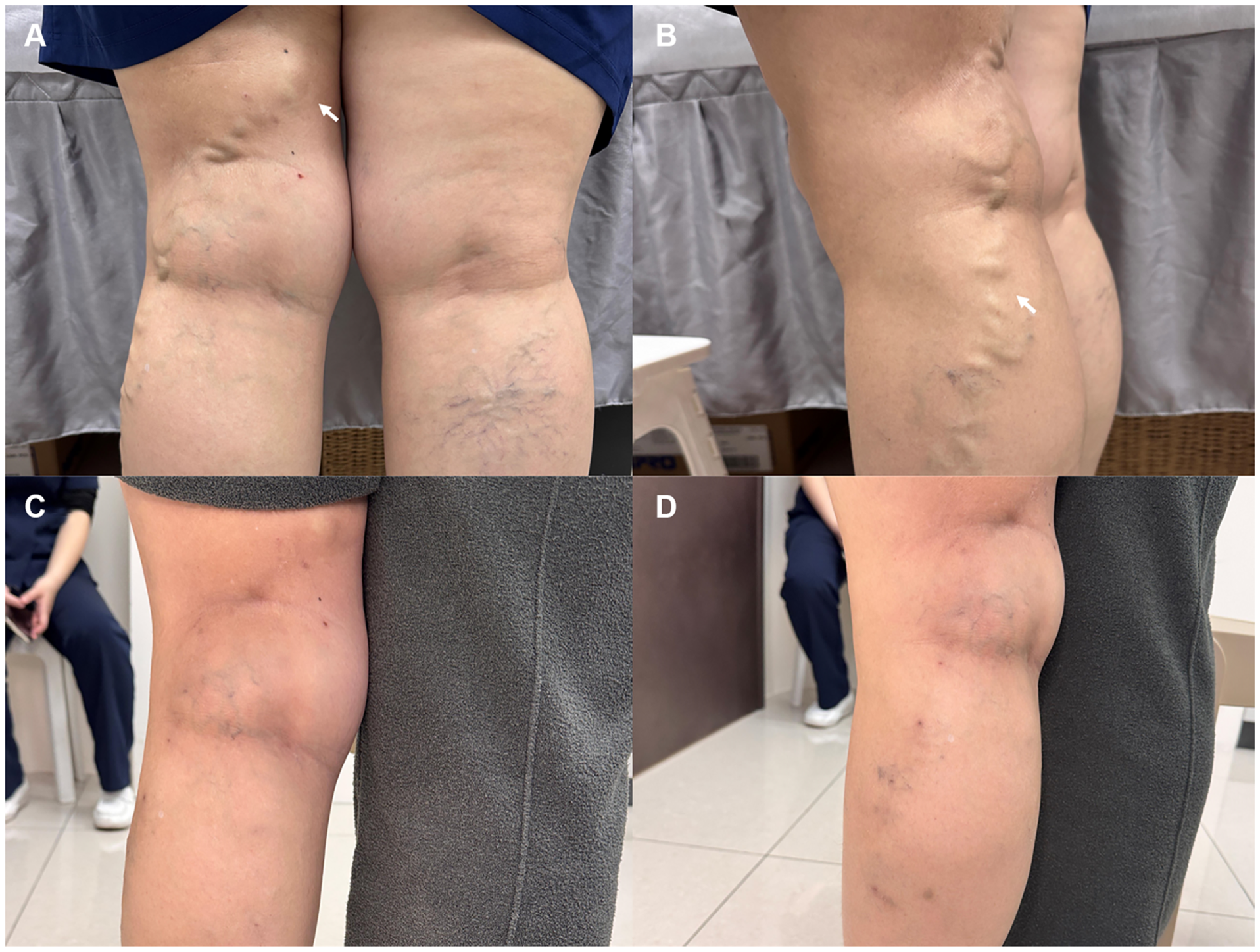
A and B show the pretreatment appearance; C and D show the three-month follow-up. The report describes resolution of the popliteal bulge and lateral varicosities. Superficial laser treatment and ambulatory phlebectomy were performed in the same session, so the appearance cannot be attributed to deep muscle branch UFLA alone. This is one patient’s outcome.
View full-size figure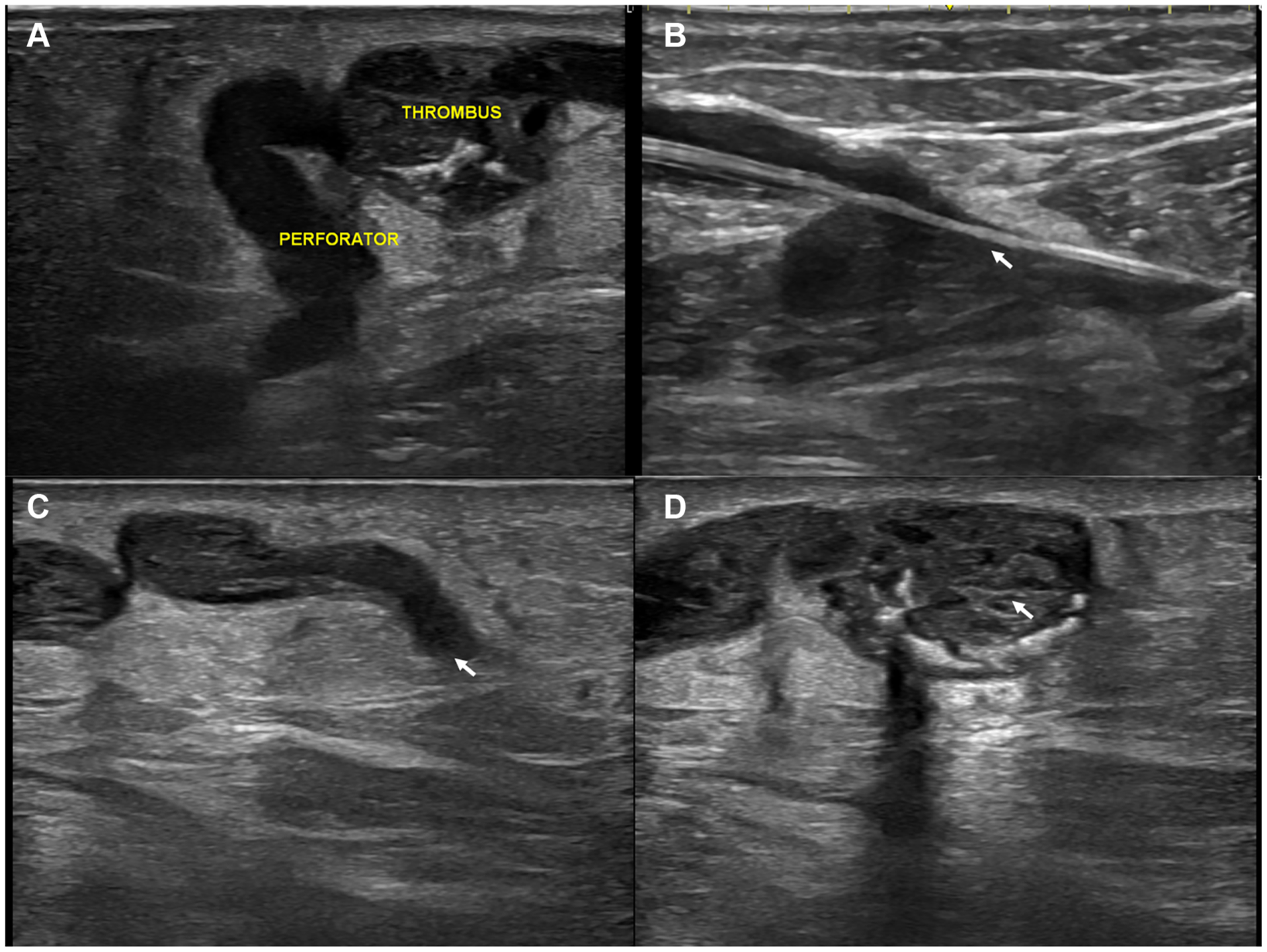
A: the refluxing branch connects to the venous aneurysm; THROMBUS denotes clot. B: a 400 μm radial fiber passes directly through an 18G catheter without an introducer sheath. C: the branch appears as a hyperechoic fibrotic cord at three months. D: the aneurysm is thrombosed, with no detectable flow. Original image labels are preserved.
View full-size figureThe BA400 R radial fiber is shown beside a standard 18G peripheral intravenous catheter. The paper reports direct passage through the catheter lumen, eliminating the introducer sheath. This is an equipment photograph, not a manufacturer’s dimensioned engineering drawing.
View full-size figureSource: Yang CC. JVS–CIT. 2026;102274. Figs. 1–3. © 2026 The Author(s). Original article
QUESTIONS & ANSWERS
Understand the principles, suitability and the limits of the evidence.
Source: Yang CC. JVS–CIT. 2026;102274. Technique, Results, Limitations Original article
Understand the source of reflux and your individual circumstances before discussing treatment.