
PRECISION IN EVERY POINT

SPOT

Segmental Point Obliteration Technique

Precision in every point.

Intermittent, point-by-point energy delivery for the below-knee great saphenous vein, exploring the balance between vein closure and protection of nearby nerves.

Chih-Chun Yang, MD · Good Leg ClinicA clinical question. A published study.

01 / THE CLINICAL CHALLENGE

Every millimeter below the knee deserves careful consideration.

Treating a refluxing vein means considering the sensory nerve beside it.

Closer than you might expect

The great saphenous vein runs along the inner leg to the saphenofemoral junction (SFJ) in the groin. In the distal calf, it can lie close to the saphenous nerve, with individual variation. Thermal ablation must balance vein closure with heat exposure to the nearby nerve.

First, understand how far reflux extends

When reflux extends below the knee, above-knee treatment alone may leave persistent distal reflux. Treatment extent depends on ultrasound, symptoms and anatomy; not everyone requires full-length ablation.
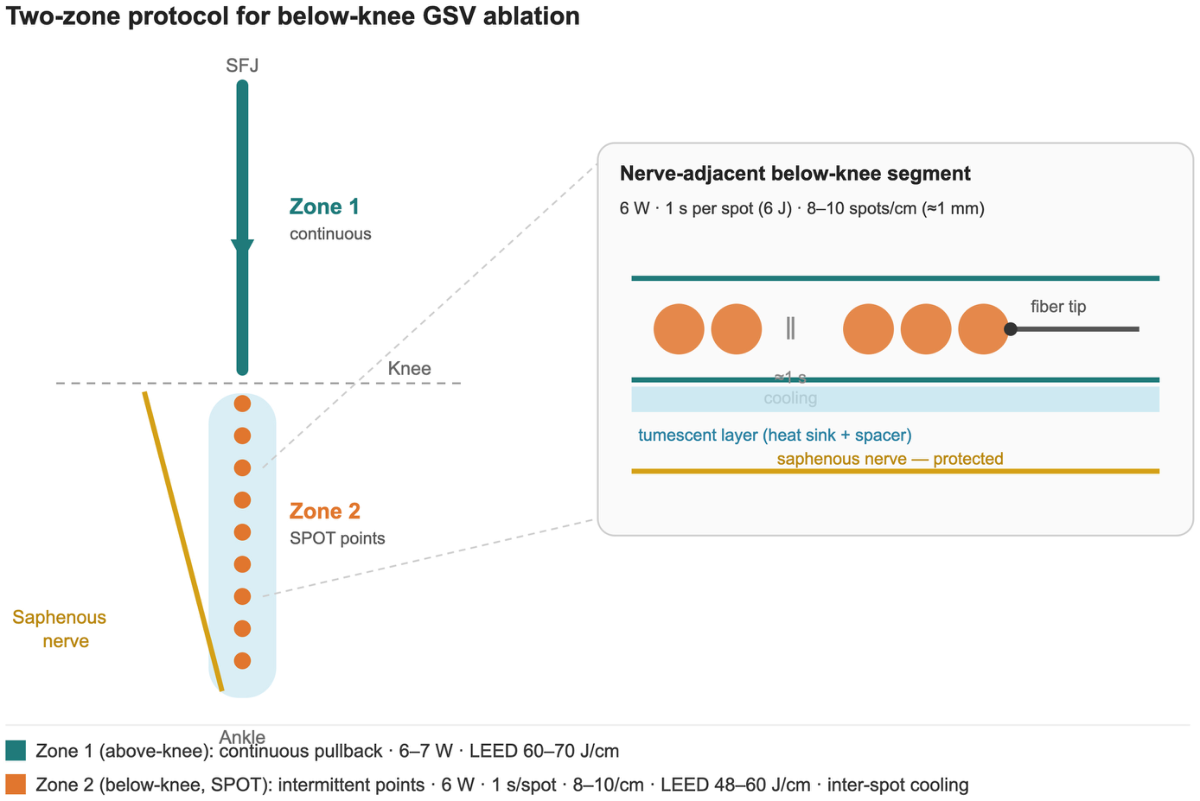
Persistent below-knee reflux is a blood-flow finding, not the overall clinical recurrence rate of varicose veins.
Fig. 1 · The two-zone protocol
Fig. 1 · The two-zone protocolZone 1 uses continuous pullback above the knee; Zone 2 uses SPOT below it. The yellow line represents the saphenous nerve. The original schematic is not anatomically to scale. “Protected” on the original figure describes the design intent, not proven freedom from nerve injury.View full-size figure

Source: Yang CC. JVS–CIT. 2026;102468. Introduction, Fig. 1 Original article

02 / THE SPOT PRINCIPLE

A different rhythm of energy delivery.

Allow time to pause and reconfirm position between treatment points.

Pulse → Withdraw → Confirm → Next point

SPOT · Sequence schematic, not a heat-spread simulation

Each point receives one second at 6 W (6 J), followed by approximately 1 mm withdrawal and ultrasound reconfirmation. Pulses are separated by approximately 1–2 seconds. Pauses are intended to permit partial cooling, while the spacing aims for contiguous treatment.

Seven points illustrate the sequence only, not a fixed treatment count or a scale drawing. The study used 8–10 points per centimeter. The link between heat dissipation and nerve protection still requires experimental verification.

Continuous energy during pullback

Zone 1 in this paper uses standard continuous pullback at 6–7 W, with LEED of 60–70 J/cm. Energy is delivered during withdrawal without inter-point pauses. This compares delivery patterns, not clinical superiority.

SPOT changes the delivery pattern rather than simply minimizing power. Energy density, power, technique and anatomy need to be interpreted together.

One vein. Two zones guided by anatomy.

The transition is determined intraoperatively where the vein lies within 5 mm of the skin and tumescent separation from the nerve becomes more difficult. It is not a fixed line at the knee joint.

Study parameters: the two-zone protocol
ParameterZone 1 · Above kneeZone 2 · Below-knee SPOT
Delivery modeStandard continuous pullbackIntermittent point pulses
Power6–7 W6 W
Linear endovenous energy density60–70 J/cm48–60 J/cm
Pulse durationContinuous1 s / 6 J
Points & withdrawal8–10 points/cm; ≈1 mm withdrawal
Inter-pulse intervalApproximately 1–2 seconds
Laser system
1470 nm · Veincare · WONTECH
Fiber selection
Maximum treated vein diameter
Maximum vein diameter <5 mm
400 μm · BA400R
Maximum vein diameter ≥5 mm
600 μm · BA600R

These settings describe the research protocol and require individual clinical judgment. SPOT does not exclusively use a 400 μm fiber, and 18G sheathless access is not the defining feature of this study.

Source: Yang CC. JVS–CIT. 2026;102468. Technique, Table I Original article

03 / EARLY CLINICAL EVIDENCE

Early findings. A starting point for further research.

January–June 2026: 13 consecutive patients with 19 symptomatic limbs, CEAP C3–C6.

13

Patients

Single operator · Single arm

19

Treated limbs

CEAP C3–C6

19/19

Complete occlusion at 1 month

Confirmed by duplex ultrasound
95% CI: 82.4%–100%

0/19

Reported paresthesia at 1 month

Saphenous nerve distribution
95% CI: 0%–17.6%

Reported paresthesia at one week was also 0/19. No deep vein thrombosis, heat-induced thrombosis, skin burn or wound infection was recorded. These are observations in a small sample, not evidence of a zero population complication rate. CI: confidence interval.

Source: Yang CC. JVS–CIT. 2026;102468. Results, Limitations, Table II Original article

04 / PRECISION IN PRACTICE

Precision begins with a complete assessment.

01

Map below-knee reflux

Assess symptoms, reflux extent, vein diameter and depth, and surrounding tissues with duplex ultrasound.

02

Tumescence & zoning

Use local tumescent anesthesia along the treatment segment. Vein depth and the difficulty of nerve separation inform the zone transition.

03

Pulse & reconfirm

Use standard continuous pullback above the knee and SPOT below it. Tip visibility is essential. Point-by-point delivery increases operator dependence and procedural time.

04

Review closure & sensation

The study assessed sensory symptoms at one week and one month, and duplex occlusion at one month. Long-term outcomes and comparative recovery remain unestablished.

What determines suitability?Symptoms, reflux extent, vein depth and diameter, adjacent tissues and overall health determine whether to treat below the knee. The fiber tip must remain visible on ultrasound; treatment should not proceed if it cannot be seen. Tumescence provides anesthesia, separation and a heat sink, but does not eliminate nerve injury.

Source: Yang CC. JVS–CIT. 2026;102468. Technique, Discussion, Limitations Original article

RESEARCH & PUBLICATION
Every claim leads back to the research.

Original journal page: Segmental point obliteration technique (SPOT) for nerve-sparing endovenous laser ablation of the below-knee great saphenous vein
SPOT2026-08-25Innovative Technique · Preliminary single-arm series

Segmental point obliteration technique (SPOT) for nerve-sparing endovenous laser ablation of the below-knee great saphenous vein

Chih-Chun Yang, MD · JVS–CIT · 13 patients, 19 limbs · 1 month

Intermittent point-by-point energy delivery was explored in the below-knee great saphenous vein. All 19 limbs were occluded at one month, with no reported saphenous paresthesia. The small sample, absent comparator and brief follow-up prevent conclusions about comparative benefit.

Journal Pre-proof (full-text version reviewed) · Journal of Vascular Surgery Cases, Innovations and Techniques
DOI: 10.1016/j.jvscit.2026.102468
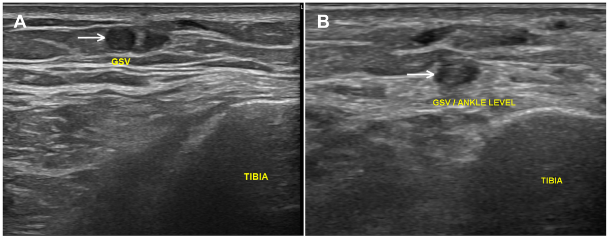
Fig. 2 · Below-knee duplex ultrasound in a representative patient
Fig. 2 · Below-knee duplex ultrasound in a representative patientAt the distal tibia/ankle level. A: a patent, round anechoic lumen before treatment. B: occlusion with loss of the anechoic lumen one month after SPOT. This is one representative patient, not a prediction for every patient.View full-size figure

Source: Yang CC. JVS–CIT. 2026;102468. Fig. 2. © 2026 The Author(s). Original article

QUESTIONS & ANSWERS

Questions deserve clear answers.

Understand the principles, suitability and the limits of the evidence.

What is SPOT?
SPOT stands for Segmental Point Obliteration Technique. Proposed by Chih-Chun Yang, MD, it is a method of below-knee great saphenous vein laser ablation using intermittent, point-by-point pulses and pauses between points. It explores a strategy for closing the vein while considering the nearby nerve.
Why do nerves need special attention below the knee?
The saphenous nerve and great saphenous vein may lie very close together in the distal calf. Heat affecting the nerve can cause tingling, abnormal sensation, or reduced sensation. Ultrasound guidance and tumescent anesthesia are important considerations, but they do not eliminate all risk.
Who may need an assessment for below-knee reflux?
People with varicose-vein symptoms, leg swelling, skin changes, or ulcers, or persistent symptoms after above-knee treatment, may need an assessment for below-knee reflux. Symptoms are not always caused by veins; a medical history, examination, and duplex ultrasound are needed to clarify the cause. This study enrolled symptomatic patients with CEAP classes C3–C6.
Does SPOT guarantee no numbness?
No. None of the 19 treated limbs had reported saphenous nerve paresthesia at one month, but the sample was small and follow-up was short. The 95% confidence interval for the event rate was 0%–17.6%. No nerve conduction studies were performed, so asymptomatic nerve changes cannot be excluded.
How does it differ from continuous laser ablation?
The difference is the delivery pattern. Continuous pullback delivers energy while the fiber moves; SPOT delivers a brief pulse at each point, followed by withdrawal and tip confirmation before the next pulse. Pauses are intended to permit partial cooling, but whether this reduces nerve injury requires larger comparative studies.
Is SPOT suitable for everyone?
No. Whether SPOT or below-knee treatment is appropriate depends on reflux extent, vein depth and diameter, surrounding tissues, symptoms, and overall health. The fiber tip must remain clearly visible on ultrasound. The paper specifically states that SPOT should not be attempted when the tip cannot be visualized.
What examinations are needed before treatment?
Lower-limb venous duplex ultrasound is central to identifying the source and extent of reflux, vein diameter, and vein depth, alongside a medical history and physical examination. The physician determines whether additional tests are needed and discusses treatment options, risks, and follow-up based on each patient’s health.

Source: Yang CC. JVS–CIT. 2026;102468. Technique, Results, Limitations Original article

Begin with a complete vascular assessment.

Understand the source of reflux and your individual circumstances before discussing treatment.