
Patient
45-year-old woman · CEAP C3
Innovative Technique · Single case report
ABSTRACT · EDITORIAL SUMMARY
A 45-year-old woman developed heaviness, swelling and a popliteal venous aneurysm 26 years after rhabdomyosarcoma resection and radiotherapy. Duplex assessment identified a 1.5 cm refluxing deep muscle branch that could not accommodate a standard introducer sheath. A 400 μm radial fiber was introduced through an 18G peripheral IV catheter; treatment used 7 W, one-second pulses and 2 mm withdrawal increments. Superficial laser treatment and phlebectomy were also performed. At three months the branch was occluded and the aneurysm thrombosed without residual flow. This single case describes short-term technical feasibility, without establishing population success rates or comparative safety.
45-year-old woman · CEAP C3
Clinical and duplex assessment
Includes superficial procedures
Observation in this case
Source: Yang CC. JVS–CIT. 2026;102274. Abstract, Results, Limitations Original article
STUDY DATA
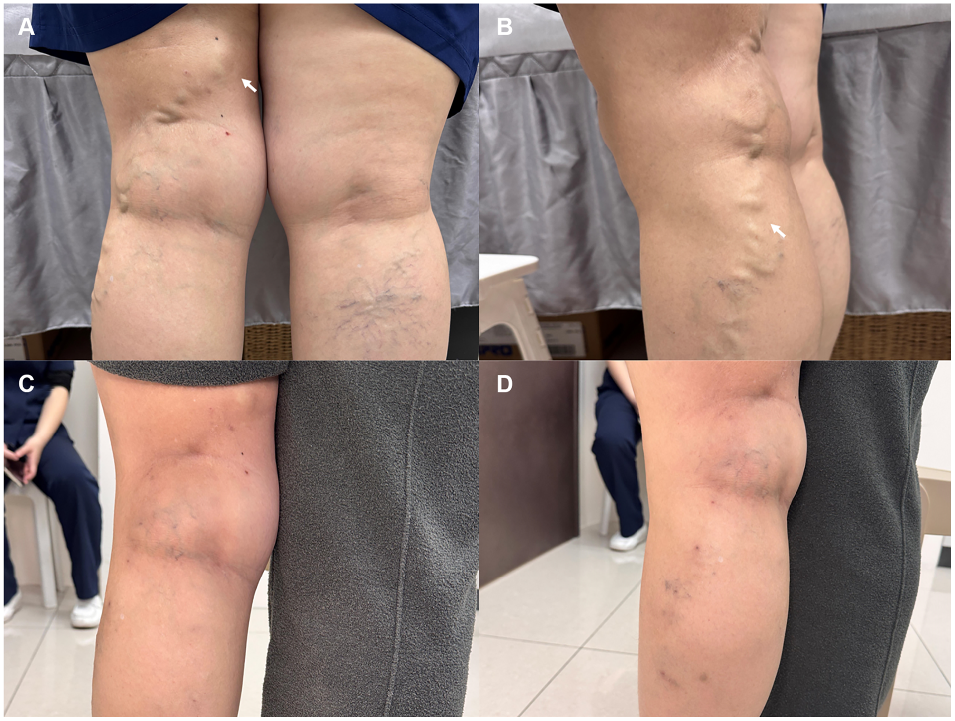
A and B show the pretreatment appearance; C and D show the three-month follow-up. The report describes resolution of the popliteal bulge and lateral varicosities. Superficial laser treatment and ambulatory phlebectomy were performed in the same session, so the appearance cannot be attributed to deep muscle branch UFLA alone. This is one patient’s outcome.
View full-size figure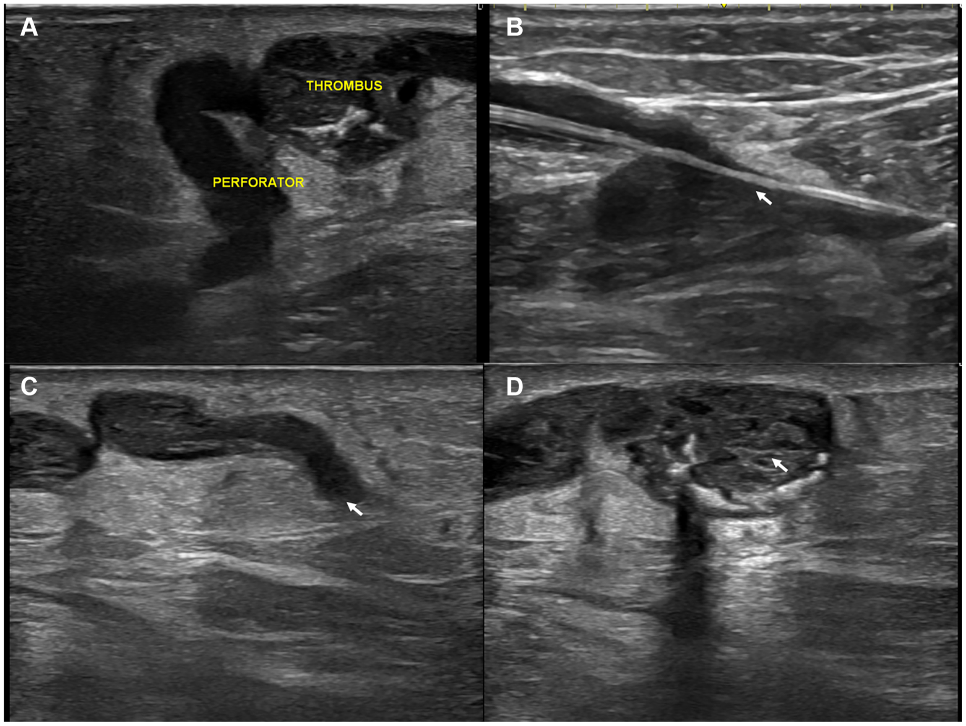
A: the refluxing branch connects to the venous aneurysm; THROMBUS denotes clot. B: a 400 μm radial fiber passes directly through an 18G catheter without an introducer sheath. C: the branch appears as a hyperechoic fibrotic cord at three months. D: the aneurysm is thrombosed, with no detectable flow. Original image labels are preserved.
View full-size figure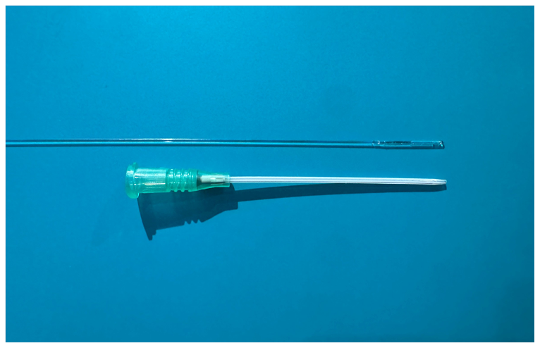
The BA400 R radial fiber is shown beside a standard 18G peripheral intravenous catheter. The paper reports direct passage through the catheter lumen, eliminating the introducer sheath. This is an equipment photograph, not a manufacturer’s dimensioned engineering drawing.
View full-size figureSource: Yang CC. JVS–CIT. 2026;102274. Figs. 1–3. © 2026 The Author(s). Original article
Source: Yang CC. JVS–CIT. 2026;102274. Figs. 1–3 Original article
CITATION
Yang CC. The sheathless ultrafine laser ablation technique for managing deep muscle branch reflux in hostile anatomy following rhabdomyosarcoma resection. J Vasc Surg Cases Innov Tech. 2026;12(4):102274. doi:10.1016/j.jvscit.2026.102274.
The UFLA online date is confirmed by PubMed; the SPOT date is from the journal-page image retained in the source project. The SPOT full text is a Journal Pre-proof; no volume or issue is inferred. Refer to the DOI for subsequent bibliographic updates.
Understand the source of reflux and your individual circumstances before discussing treatment.